
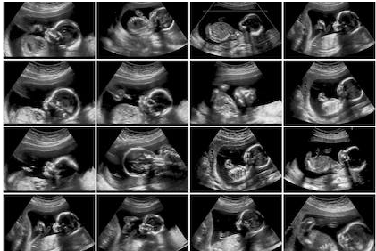
A dating scan is an ultrasound scan done between 8 and 14 weeks of pregnancy to help estimate your baby’s due date. In early pregnancy, most babies of the same gestational age are about the same size.
A dating scan measures your baby and this helps your doctor estimate how long you have been pregnant, and when your baby is due.
Not everyone needs a dating scan, but they can be very helpful if you aren’t sure when you conceived.
It’s important to have an accurate estimated due date (or EDD) for your baby so you can have the recommended tests at the right time.
Knowing how far along you are is also important if your baby is born prematurely, or if you haven’t given birth by your
estimated due date and you’re thinking about having your labour induced.
Most babies are born about 38 weeks after conception. Since many women ovulate (release an egg that may then be fertilised)
and conceive about 2 weeks after their last period, this is often about 40 weeks since the beginning of their last period. That’s why people often talk about pregnancy lasting for 40 weeks.
Women with a regular 28-day cycle can calculate an estimated due date for their baby by counting 40 weeks from the first day
of their last menstrual period. This may not be so simple or accurate in other situations, like if you have long or irregular cycles,
don’t remember when you had your last period, or if you got pregnant while taking contraception that affected your cycle.
If you think you may need a dating scan to help estimate when your baby is due, speak to your doctor or midwife.
A dating scan is done between 8 and 14 weeks of pregnancy (and usually between 8 and 12 weeks), when most babies of the same gestational age are about the same size. If your doctor or midwife thinks you should have a dating scan as well as a test for nuchal translucency, they may recommend you arrange it between 11 and 13 weeks, so you can have both tests during a single ultrasound scan.
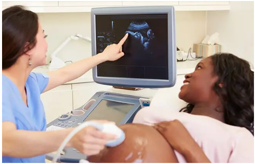
A dating scan is usually performed by a specially-trained technician called a sonographer, but it can also be performed by anyone who is trained to do it. This may include doctors, midwives or other health workers. It may be performed in a radiology clinic or a hospital.
In early pregnancy, ultrasounds including dating scans can be done through your abdomen (tummy) or vagina.
The method used will depend on a few factors, including how far along your pregnancy is and your body shape.
If your scan is being done along your abdomen (known as a ‘trans abdominal ultrasound’), you will be asked to
drink a few cups of water before you arrive so your bladder is full. This can make it easier to see inside your uterus (womb).
The sonographer will apply some gel and gently move the ultrasound probe along your abdomen. It doesn’t usually hurt.
If your scan is done through your vagina (known as a ‘transvaginal ultrasound’),
a small ultrasound probe is lubricated and gently inserted into your vagina.
The probe may be a little uncomfortable but usually isn’t painful. Scans done this way can give more detailed pictures because the probe is closer to your uterus.
Ultrasounds, including dating scans, do not harm you or your baby or increase your risk of miscarriage.
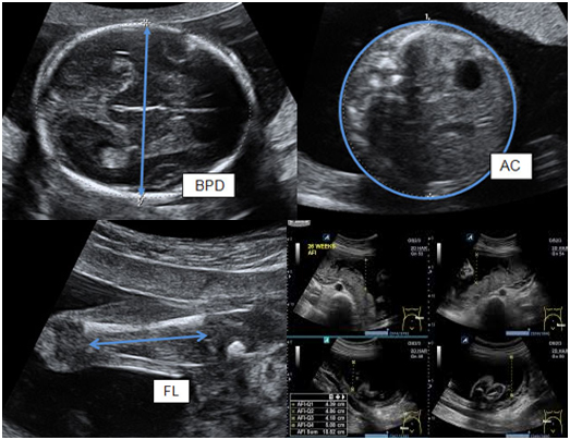
During the scan, the sonographer will measure your baby’s length from head to bottom, known as their ‘crown-rump length’ (CRL).
This measurement can help estimate your baby’s gestational age and when it is likely to be born.
Having an accurate estimated due date is helpful, but it’s also important to remember it’s only an estimate.
Most babies are not born on their due date.
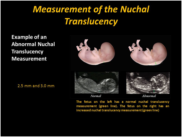
A nuchal translucency scan is an ultrasound scan that measures your baby’s nuchal translucency — a fluid-filled
space behind your baby’s neck. This measurement can help your doctor estimate the risk of your baby having a chromosomal
abnormality such as Down syndrome..
The chances of having a baby with a chromosomal abnormality are greater the older you are when
you get pregnant. However, anyone can have a baby with chromosomal abnormalities, so screening is
offered to everyone, but the decision to have the scan is yours.
While scans can reassure you that your baby is developing normally, you may also learn that your baby has an
abnormality. For this reason, before you have the test it’s a good idea to think about why you are choosing to do it,
and what you would do next if your screening test showed you were at a high risk of your baby having a chromosomal abnormality.
Some women choose not to have any tests or decide to have a diagnostic test instead (such as chorionic villus sampling or amniocentesis), which can give them more definite information about their baby’s health.
A nuchal translucency scan is done between 11 and 14 weeks of pregnancy. If your doctor has referred you for a dating scan, it can often happen at the same time.
A nuchal translucency scan is usually performed by a specially-trained technician called a sonographer, but other health professionals, such as doctors or midwives with the relevant training, may also do it. The scan may be performed in a radiology clinic or a hospital.
In early pregnancy, ultrasounds such as the nuchal translucency scan can be done through your abdomen or vagina.
Your sonographer will choose a method based on several considerations, including how far along your pregnancy is and your body shape.
If your scan is done along your abdomen — trans abdominally — you will be asked to drink a few cups of
water before you arrive so that your bladder is full. This makes it easier to see inside your uterus (womb).
The sonographer will apply some gel and gently move the ultrasound probe across your abdomen. It doesn’t usually hurt.
If your scan is done transvaginally, a small, lubricated ultrasound probe is gently inserted into your vagina.
The probe may be a little uncomfortable but usually isn’t painful. Scans done this way can give more detailed pictures because the probe is closer to your uterus.
Ultrasounds do not harm you or your baby or increase your risk of miscarriage.
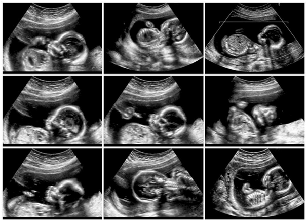
The target scan is one of the most important prenatal ultrasound scans.
This scan is performed to assess the fetus’s development and location, as well as to detect any impairments.
This scan aids in the detection of anomalies and, in certain extreme cases, allows you to decide whether to continue the pregnancy.
As with many pregnancy exams and treatments, this one is also a personal choice for the woman. However, all prenatal experts strongly suggest this scan.
The target scan is done between 18 and 20 weeks of pregnancy because the baby grows around six inches by the 19th week of pregnancy. Several anatomical structures and internal organs begin to develop and mature, so the target scan captures any abnormalities in the fetus.
Although many abnormalities are not detectable during sonography, about 50 percent of Down syndrome problems and congenital heart defects are identified.
The target scan evaluates the fetus from the head to toe, checking general growth, development, and health. The sonographer evaluates the baby’s anatomy and any structural issues.
The scan can determine the fetus’s size and weight. Moreover, it considers the location of your placenta, umbilical cord, and amniotic fluid surrounding the fetus.
Any improper location of the placenta during pregnancy can result in miscarriages or severe bleeding, as well as a variety of issues for both the mother and infant.
In certain circumstances, the baby’s organs do not grow normally. Some may result in the death of the fetus anytime during the pregnancy or shortly after birth. All of this must be discovered, investigated, and analyzed beforehand. This scan allows your obstetrician to provide the appropriate steps, medications,
and reassurance about your baby’s health for the remaining days of your pregnancy.
A morphology (body part) scan is a routine antenatal test usually done at 18 to 20 weeks of pregnancy. It is an ultrasound that checks your baby’s size and body organs. Your doctor is likely to recommend you have this test, but the decision to do so is yours. A morphology scan is sometimes called a ‘fetal anomaly’ scan since it is one way your doctor will check your baby for birth defects
The scan is usually done by our ultrasound technician (sonographer) who is specially trained to check your baby. Sometimes one of the clinic’s specialist doctors may also come in to scan your baby to help get as much information as possible.
Some clinics will ask you to drink 3 glasses of water 1 hour before the appointment and to then hold it in. This is because having a full bladder makes it easier to see the images. Other clinics recommend eating and drinking as normal, but ask that you do not empty your bladder within 30 minutes of your appointment. Please check what the clinic prefers when you book your appointment.
The sonographer will place some gel on your abdomen (tummy) before using a probe on your skin, known as a transducer, to create images of your baby. Pulses of sound waves will move from the transducer to bounce off your baby, creating echoes. The computer then changes these echoes into images.
The sound waves are harmless to your baby and they will be unable to hear the scan as the volume is very low.
The results of the scan will be available on the same day that you have it. A copy of the report will go to your referring doctor. If there are any abnormalities found during the scan, a specialist doctor will contact you to discuss what they mean.
If the baby has an average head size, but a big abdomen, he/she may just be getting a good food supply from the placenta. If the mother has diabetes in pregnancy (gestational diabetes) she may have a big baby (macrosomia). This happens when the mother’s blood sugar levels have risen too high. This can make the baby measure larger than expected for all parameters.
If the baby has an average head size and a small abdomen,, this may simply indicate a small, healthy baby. Occasionally, though, this can be a sign that the baby is not growing properly. The scan may also show that the amount of amniotic fluid is low. The two sometimes occur together.
If the baby is smaller than expected, he/she may have a low birth weight. To find out why the baby is small, the doctor might ask for a Doppler scan.
Scans are usually accurate for assessing the baby’s size in the first half of the pregnancy. By the time the mother is in the later stages of pregnancy, scans continue to be accurate, as long as the baby is small or of average size. The closer it gets to the due date, and the bigger the baby is, the harder it will be to record measurements.
The baby’s head may be too low in the pelvis in late pregnancy to get a measurement. Even if the baby’s abdomen can be measured, it’s very difficult to take other factors into account, such as how long the baby is.
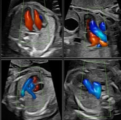
In Nuvha Scan, Fetal echocardiography is a test similar to an ultrasound. This exam allows your doctor to better see
the structure and function of your unborn child’s heart. It’s typically done in the second trimester, between weeks 18 to 24.
The exam uses sound waves that “echo” off the structures of the fetus’s heart.
A machine analyzes these sound waves and creates a picture, or echocardiogram, of their heart’s interior.
This image provides information on how your baby’s heart formed and whether it’s working properly.
It also enables your doctor to see the blood flow through the fetus’s heart.
This in-depth look allows your doctor to find any abnormalities in the baby’s blood flow or heartbeat.